Dental X-Rays

Dental X-rays can detect problems that would be missed by just looking in your mouth
▪an infection in your tooth or tooth root;
▪cavities between teeth or under fillings;
▪trouble with teeth and jaw development in children and teens who are getting their permanent teeth;
▪bone loss from severe gum disease.
Sometimes x-rays are needed as part of your dental treatment. When deciding whether to recommend x-rays, your dentist will consider factors such as
▪your current oral health, including any oral health problems you are having;
▪your age;
▪your risk for tooth decay or gum disease.
Your dentist might recommend x-rays if you are a new patient. X-rays help your dentist evaluate your oral health and give him or her something to compare against when looking at changes that may occur later.
Common Types of Dental X-Rays
There are several types of dental x-rays. Each one helps the dentist see different areas of your mouth.
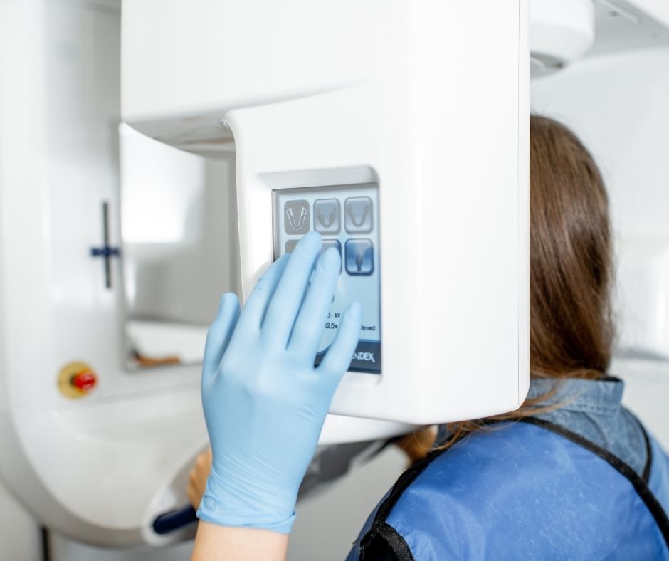
Common x-rays used in the dental office include bite-wing, periapical, and panoramic x-rays. Bite-wing x-rays help the dentist check for tooth decay between the back teeth or under dental fillings. These are taken with a small film or digital sensor that you bite with your back teeth. Periapical x-rays help the dentist observe conditions below the gumline, showing the roots of the teeth. They can be taken of front or back teeth. Panoramic x-rays use a machine that rotates around the head. It produces a long film that shows the entire jaw and all of the teeth in 1 image.
Cone-beam computed technology is used to make a special type of x-ray that is used less often. This technology takes a series of images to create a 3-dimensional image. Because it relies on multiple images, the radiation exposure is higher than that of commonly used x-rays. It is used when more detailed information is needed.